JOINTS 2024;
2: e1353
DOI: 10.26355/joints_202412_1353
Pseudoaneurysm of the superior medial geniculate artery after total knee arthroplasty
Topic: Knee
Category: Case report
Abstract
BACKGROUND: The incidence of vascular injuries is reported to be low after total knee arthroplasty (TKA). Among them, pseudoaneurysms of geniculate arteries (GAs) are considered to be a rare complication. The pathogenetic mechanism to develop this complication usually involves iatrogenic damage of GAs that may potentially occur in any phase of the surgical procedure. The diagnosis of pseudoaneurysm lesions of GAs could be delayed, negatively affecting postoperative rehabilitation. From a clinical viewpoint, a pseudoaneurysm around the knee could determine a pulsatile mass, pain, swelling and reduced articular function. Despite there is not a gold standard for the diagnosis, different imaging methods such as ultrasound (US), computer tomography (CT) and magnetic resonance imaging (MRI) could play a role.
CASE REPORT: The aim of our study is to report a case of a male patient who developed a pseudoaneurysm of the superior medial geniculate artery after TKA, describing the clinical and radiological presentation and the diagnostic and therapeutic pathway and comparing our findings with literature.
CONCLUSIONS: Despite pseudoaneurysms of the geniculate arteries being a rare complication after TKA, it is possible to suspect that its incidence may be higher than reported in the literature because not all cases are clinically evident. Furthermore, the characteristic clinical features that lead to suspect the diagnosis, such as a palpable pulsatile mass, are reported to be not present in all cases, and there is not a typical range of time for the clinical onset. The diagnosis is insidious and should be suspected, especially in patients with a swollen and painful knee with risk factors for this complication. A prompt diagnosis is crucial to prevent the delay of the rehabilitation program and to improve functional recovery. Ultrasound is the most appropriate imaging method for a primary evaluation, as it shows characteristic hallmarks such as the “yin-yang” sign. A second-level imaging evaluation is needed to confirm the diagnosis, but the appropriate radiologic method is still being debated. The choice between CT and MRI should be evaluated with an interdisciplinary approach and according to their ready availability. The interventional angiography with embolization procedure is considered safe and effective to resolve the pathology.
Introduction
The development of arterial pseudoaneurysms is rare after total knee arthroplasty (TKA), with an incidence ranging from 0.03% to 0.2%1,2. Despite studies that have reported the development of this complication after TKA3-5, other common causes of arterial pseudoaneurysms after knee surgery are open synovectomy, meniscectomy, trauma, infections, and invasive intravascular diagnostic procedures6-8. The most common localization of pseudoaneurysms around the knee is the popliteal artery, followed by the superolateral, inferolateral and inferomedial geniculate arteries (GAs)8. When considering the TKA surgical procedure, an injury of the popliteal artery can occur in knees with flexion contracture during the posterior capsule release manoeuvres9, while the geniculate arteries could be injured, especially during the soft-tissues balancing release procedures on the collateral ligaments3. The incidence of this complication is increased in patients with fracture sequelae or in patients with atherosclerotic arteries because of arterial abnormal anatomy or reduced arterial wall elasticity, respectively2,9. Despite literature reporting the development of an arterial pseudoaneurysm as a rare complication after TKA, it is possible that the epidemiology of this complication, in particular regarding GAs, is underestimated and only a percentage of artery injuries can be detected and managed intra-operatively and only a percentage can results clinically evident. Usually, a pseudoaneurysm causes pain, reduced range of motion (ROM), recurrent hemarthrosis, and swelling, and only sometimes, it may present as a pulsatile mass. Furthermore, it impairs knee function, and consequently, patients have delayed postoperative rehabilitation. In other cases, it may determine sensitive or motor deficit and decreased peripheral arterial pulse5,8,10.
The diagnosis of this complication is often delayed and requires a high level of suspicion and a combination of clinical and radiological findings. Plain radiographs are not as useful as ultrasound (US), computer tomography (CT), and magnetic resonance imaging (MRI)4,11.
The aim of our research is to report a case of a patient who developed a pseudoaneurysm of the superior medial GA after TKA and to describe our diagnostic and therapeutic pathway.
Case Presentation
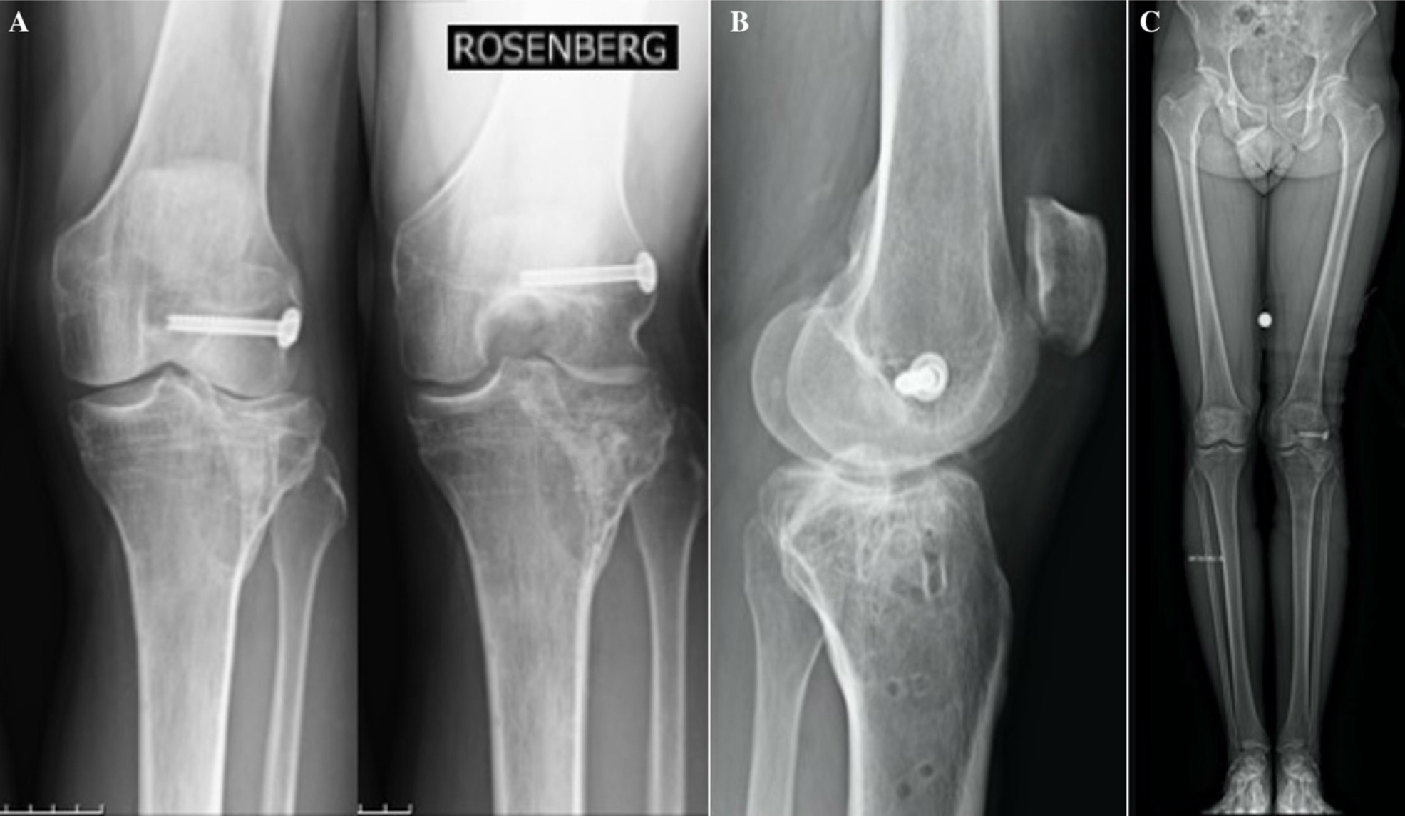
A 41-year-old male patient with a 5° coronal valgus deformity of the left knee and severe osteoarthrosis (Kellgren-Lawrence grade 4) underwent TKA. The patient had a history of a lateral tibial plateau fracture associated with lateral collateral ligament (LCL) lesions, which were both surgically managed in other institutions with open reduction and internal fixation (ORIF) combined with LCL repair, respectively (Figures 1 and 2).
Figure 1. Preoperative X-ray. Anteroposterior preoperative X-ray of the left knee with Rosenberg view (A) and lateral view (B) showing severe osteoarthrosis in the setting of a previous lateral tibial plateau fracture, managed with open reduction and internal fixation. Lateral collateral ligament rupture was treated with a metallic fixation screw, still visible. Weight-bearing radiographs of the lower extremities (C) show coronal valgus deformity on the left side.
The patient had no medical comorbidities. The surgical procedure of TKA was performed using a mini-mid vastus approach and a kinematical alignment technique without a tourniquet. A mobile-bearing cemented TKA (Innex total knee, Zimmer-Biomet, Warsaw, IN, USA) was implanted in 45 minutes (Figure 3). During the surgical approach and after the prosthesis implantation, before proceeding with the suture, an evaluation of active hemorrhages and normal hemostasis procedures for small vessels was performed using the monopolar electrosurgical instrument. At the end of surgery, there was no evidence of active macroscopic hemorrhages or main vessel injuries. Despite this, an intra-articular drain was positioned as our standard protocol after joint replacement surgery and removed 12 hours after surgery, containing 200 ml of blood. After surgery, the patient underwent our standard post-operative medical care protocol based on analgesic medications (paracetamol 1 g twice a day and morphine administered consecutively for 48 hours by using an elastomeric pump), non-steroidal anti-inflammatory drugs (ibuprofen 600 mg twice a day), anti-thromboembolic prophylaxis (nadroparin calcium weight-dosed) and cryo-therapy (local ice pack 3 times a day). The patient had a normal postoperative recovery, and no blood transfusions were necessary. On postoperative day 5, he was discharged and transferred to a rehabilitation hospital. At discharge, his hemoglobin was 10.5 g/dl, the pain was controlled by standard analgesic medications, and the knee swelling was consistent with the recent surgery.
Figure 3. Postoperative weight-bearing radiographs. Postoperative weight-bearing radiographs (A) showing restoration of the regular femoral-tibial load axis. B-D, show the regular positioning of the mobile bearing cemented total knee arthroplasty.

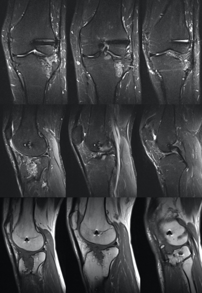
On postoperative day 15, the patient developed increased left leg swelling and increased pain, and he returned to the emergency department of our institution. On clinical examination, he presented a palpable mass in the medial portion of the distal region of the thigh, proximally to the medial femoral condyle. He had no neurological symptoms, but his blood exams showed a stable hemoglobin value of 10.5 g/dl. Inside the emergency department, a diagnostic arthrocentesis was performed, removing 50 cc of blood; the sample was sent to our microbiology department to check for infection, but the result was negative. Then, a US evaluation of the knee was performed by an expert radiologist. The US examination showed an inhomogeneous joint fluid collection in the suprapatellar space, with hyperechoic internal echoes, suggesting the presence of hemarthrosis. When moving the probe to the superomedial side of the knee, a small hypo-anechoic rounded mass was detected. The color-Doppler evaluation showed a turbulent flow within the lesion, with a characteristic “yin-yang” sign due to the forward and backward flow that led us to suspect the diagnosis of supero-medial artery pseudo-aneurysm (Figure 4).
Then, a computed tomography angiography (CTA) was performed, confirming the diagnosis of pseudoaneurysm of the superomedial geniculate artery (Figure 5). The maximum diameter of the pseudoaneurysm was 15 mm (transverse) and 14 mm (longitudinal). Moreover, the pseudoaneurysm was surrounded by a large hematoma (maximum transverse diameter of 6 cm).
Figure 4. Ultrasound evaluation of the knee. A, A large, inhomogeneous suprapatellar effusion (solid arrow) is visible, characterized by internal echoes but with no color signals within or around on power-Doppler, suggesting hemarthrosis. B, At the superomedial side of the knee, a round formation (dashed arrow) with turbulent flow on color-Doppler is shown, displaying the characteristic “yin-yang” sign. This formation is surrounded by a hematoma (*). A vessel with turbulent flow (dashed arrows) crosses the formation. Both (B) and (C) show the presence of hemarthrosis # deep in the vascular formation.

Figure 5. Computed tomography angiography of the knee. Not-enhanced CT shows a slightly hyperdense collection on the anteromedial region side of the knee (hemorrhage), with an internal rounded hypodense area (*). After iodine-contrast administration (B), the rounded area turned out to be a contrast-enhancing vascular structure, confirming the diagnosis of pseudoaneurysm (arrow). C (axial) and D (sagittal) show CT maximum intensity projection (MIP) that clearly depicts the origin of the pseudoaneurysm (arrow) from the superomedial geniculate artery (dashed arrow).

The patient was transferred to the emergency department of the Niguarda Hospital (Milan, Italy) and carried in the interventional radiology (IR) suite. Under local anesthesia, a 5 French sheath was placed with ultrasound assistance in the left common femoral artery through an antegrade approach. The arteriography of the limb confirmed the presence of a pseudoaneurysm originating from the medial superior genicular artery; in particular, it was a saccular pseudoaneurysm associated with iatrogenic/vasospastic stenosis immediately before its neck (Figure 6A-B). A 4 French catheter was advanced until the origin of the medial superior genicular artery (Figure 6C). Then, a 2.7 French coaxial micro-catheter and a 0.021″ guidewire were used to catheterize the vessel just beyond the origin of the pseudoaneurysm. Two detachable coils (2 mm x 4 cm; Concerto, Medtronic, Minneapolis, MN, USA) were placed from this site going backward to the neck of the pseudoaneurysm; then, other three pushable coils (3 mm x 4 cm and 3 mm x 8 cm; Nester, Cook Medical, Bloomington, IN, USA) were placed going backward to embolize the artery just before the origin of the pseudoaneurysm. The final angiography confirmed the complete embolization of the pseudoaneurysm and the preserved patency of the proximal tract of the medial superior genicular artery (Figure 6D). The femoral access was then closed with a mechanical device (Angio-Seal VIP, Terumo Europe, Leuven, Belgium).
Four days after the embolization, a follow-up ultrasound showed the pseudoaneurysm filled with hyperechoic material (metallic coils) and the absence of vascularization. Both the hemarthrosis and the soft tissue hematoma were reduced (Figure 6E).
Figure 6. Interventional angiography. A-B, show the angiography on two subsequent frames, highlighting the presence of the pseudoaneurysm originating from the medial superior genicular artery (arrow), with progressive filling. C, shows the selective angiography from the medial superior genicular artery, confirming the saccular pseudoaneurysm. Post-procedural final control (D) shows the complete embolization of the pseudoaneurysm by coils (arrow). E, shows the follow-up ultrasound after four days with the pseudoaneurysm filled with hyperechoic material (coils) and the absence of vascularization at color-Doppler.

Following the embolization procedure, the patient returned to our institution. After 24 hours of rest and combined compressive cryotherapy, he started intensive physiotherapy to recover the full ROM of the knee. Then, two days after the embolization procedure, he was discharged at home. At the time of discharge, the knee ROM was 0-110°, and the patient was able to walk with two crutches and full weight-bearing. Moreover, his hemoglobin was increased to 2.2 g/dl.
Discussion
Literature Review
A literature review was performed. The following databases have been utilized: PubMed, Google Scholar, Embase, and Cochrane. The keywords used were ‘geniculate arteries injury,’ ‘geniculate artery pseudoaneurysm,’ ‘vascular lesions,’ ‘vascular injuries’ associated with ‘TKA’, and ‘total knee arthroplasty.’ Only articles in the English language were considered. Since the low volume of literature on the topic, articles were screened for eligibility after a careful reading of the abstract. Cross-reference research of the selected articles was performed to find other relevant papers on the topic. Different article types have been included in the final review: meta-analysis, systematic reviews, case report case series, and retrospective studies.
Epidemiology
Complications involving vascular injuries are infrequently reported after total knee arthroplasty (TKA) and are considered rare complications. A large systematic review and meta-analysis performed by Sundaram et al12 confirmed the low risk of vascular injuries associated with TKA, reporting 767 cases that occurred in 1,419,557 knee arthroplasties, corresponding to 0.054%. This is confirmed by a recent retrospective case series study13 that found an incidence of vascular lesions after TKA of 0.088%.
Regarding pseudoaneurysm, Hodgson et al14, in their systematic review, reported a prevalence of 0.6%.
Even if the iatrogenic damage of the geniculate arteries is mostly considered in literature a rare occurrence that is often already assessed and managed intra-operatively, it is reasonable to hypothesize that minor damage of these arteries can occur more frequently than reported in literature and only a percentage of these patients can show the typical clinical features related to this complication. Daniels et al4, in their retrospective study including only symptomatic pseudoaneurysm after TKA diagnosed on imaging, confirmed the low incidence of this complication at approximately 0.02% but hypothesized a higher incidence considering that some may have been diagnosed clinically without imaging confirmation, and some small pseudoaneurysms may go undetected at clinical and radiologic evaluation.
The specific rate of injury to smaller periarticular vessels around the knee has not been well studied in the literature, and more studies focusing on the incidence of geniculate artery pseudoaneurysm after TKA are needed to determine the real entity of this complication.
Pathogenesis and Injury Mechanism
GAs injuries may potentially occur in any phase of the surgical procedure. These arteries are vulnerable during dissection, during femoral and tibia cuts, as well as during retraction due to the blunt trauma from instrumentations, such as retractors12, and during the soft-tissue balancing release procedures on the collateral ligaments.
Pseudoaneurysmal lesions of GAs are reported to involve more frequent lateral GAs than the medial GAs4.
Literature shows that GAs are at risk for injury during specific surgical gestures associated with TKA. The superior-lateral geniculate artery is at risk for injury during lateral retinacular release, while the inferior-lateral GA is vulnerable during dissection, excision, cutting of the tibia, and cutting near the lateral meniscus, as well as while retracting soft tissue from the tibial edge, especially in mini-invasive surgery5,12,15-17. The inferior medial GA is at risk for injury during the release of the medial structures for soft-tissue balancing and during cuts near the medial meniscus3,12. The superomedial GA injury could be due to excessive knee extension and flexion during TKA placement5,18 and during medial soft tissue release in the attempt to correct a varus deformity4.
Clinical Features
Patients with a pseudoaneurysm of a GA could be asymptomatic or may present different symptoms, in most cases not specific to this kind of complication. Several studies12,19 show how the first step to reaching the diagnosis is to think about this complication; this diagnosis requires being clinically suspected with high clinical awareness and a watchful clinical attitude in front of painful and swelling knees after surgery, especially in patients considered at high risk for these complications. This watchful approach includes investigating preoperatory history, focusing on potential risk factors reported to be previous knee surgery or fractures, diabetes, peripheral vascular disease, hypertension, and smoking status12,13.
Sundaram et al12 proposed a perioperative management algorithm that categorizes patients into high or low risk based on risk factors for vascular injuries. Post-operative frequent physical examinations are required in high-risk patients, with ultrasound being the first imaging modality in injury-suspected cases. The most common clinical presentation of a pseudoaneurysm after TKA is a painful and swollen knee with recurrent hemarthrosis and a decreased range of motion with a detectable mass that, only in a minority of cases, can show a characteristic pulsatility4,12,18,20-22. Furthermore, anemia could be found in blood exams.
While most case reports and case series3,5,8,10,20 in the literature agree on the main features of clinical presentation, different data are reported regarding the timing of clinical onset. In our case, symptoms appeared on the 15th day postoperatively; however, the literature8,12,14,19,21,22 shows that the timing of clinical onset can range from a few days to several months after surgery.
Sundaram et al12, in their meta-analysis, reported 18 cases of GAs pseudoaneurysm, all diagnosed after 24 h. Other works in the literature report different and delayed timing of clinical onset after surgery.
A systematic review performed by Hodgson et al14 reported 3.5 days as the average time taken to reach the diagnosis of pseudoaneurysm after TKA.
Furthermore, several case reports describe the possibility of delayed clinical onset and pseudoaneurysms diagnosed later after surgery, from a few days19,21 to several weeks8 or months22.
Probably, this difference concerning the clinical onset may be due to patient-specific anatomic local conditions and to the progressive pathogenetic mechanism by which the pseudoaneurysm develops starting from the presence of an arterial wall lesion until the main clinical features can emerge.
Radiologic Evaluation
Literature4 indicates that there is no definitive gold standard for diagnosing knee artery pseudoaneurysms, and various radiologic examinations may be significant in this process. The available literature4,5,12,18,19,21 suggests ultrasound (US) may be considered the most appropriate primary imaging method, allowing a rapid evaluation of TKA-associated vascular injuries. In most cases, it can be diagnostic and effective, presenting characteristic findings such as a lobate mass with varying echogenicity next to the parent artery. This mass exhibits bidirectional color Doppler flow with a swirling motion, creating the distinctive “yin-yang” sign in color Doppler imaging ultrasound4. According to this, our diagnostic work-up showed the effectiveness of US imaging performed by an expert radiologist in the initial imaging evaluation; it aroused significant suspicion regarding the diagnosis, particularly upon discovering the typical hallmark known as the “yin-yang” sign.
Despite this and according to the literature4,5,12,18,19,21, we deemed it necessary to conduct a second-level instrumental investigation in order to confirm the diagnosis and direct the patient to the correct treatment.
The selection of the following second-level imaging exam is debated in the literature4,11 and should be evaluated with an interdisciplinary approach with colleagues in vascular surgery and/or interventional radiology, considering the possibility of treatment and the availability of the different options12.
Daniels et al4, in their work, consider MRI/magnetic resonance angiography (MRA) as the most appropriate second-level exam; in particular, even if MRI could be useful, MRA could be considered more effective, identifying the pseudoaneurysm and the parent artery as a mass in close proximity to the artery that rapidly enhances in the arterial phase and washes out in the late venous phase4. It offers advantages in terms of radiation exposure and is considered the examination of choice for a painful TKA, even if it remains a time-consuming technique with limited availability if compared with US or CT, especially in trauma settings and emergency departments11.
For these reasons, other authors support the role of computed tomography angiography (CTA)11, although it requires a high radiation dose with the possibility of a high false-negative result4.
Conventional angiography can confirm the presence of pseudoaneurysm in most cases and also plays an important role in treatment4.
Daniels et al4 propose an imaging algorithm for the workup of a painful and swollen knee after TKA: US is a good initial test because it is fast, cheap, and does not require intravenous contrast medium or radiation. MRI with MRA should be performed if other abnormalities need to be assessed in addition to a pseudoaneurysm or if ultrasound is negative with high clinical suspicion for a pseudoaneurysm.
In our case, after US evaluation and according to the radiologist, we preferred to proceed with a second-level imaging exam to confirm the diagnosis; we opted for a CTA scan, both because this technique is rapid and immediately available in our emergency department setting (as the event happened on a public holiday).
Treatment
Most of the literature12,13 reports endovascular treatment as the most common, safe, and effective solution for TKA vascular injuries. This treatment is mini-invasive and allows for rapid clinical improvement in most patients; open surgery has been confined to specific cases where direct repair or bypass is required.
According to this, our case report describes the treatment, which took place in an interventional radiology suite with endovascular coil embolization. It has been shown to be effective, without any complications, and without important interference with the rehabilitation program after TKA.
Only a few works in the literature describe the alternative possibility of non-operative treatment for GA pseudoaneurysms after TKA. Sundaram et al’s12 meta-analysis reported only 2 cases of non-operative treatment, while Gaheer et al18 described a case report of a spontaneous resolution of a superior medial GA pseudoaneurysm.
Angiography is a tool of great diagnostic and therapeutic value13. This procedure is safe, with a lower complication rate and a lower risk of infection than an open procedure. It is performed under local anesthesia, allowing diagnosis and treatment during the same procedure and a quick return to the patient’s rehabilitation program19,23,24.
Analyzing several case reports and series19,17,21, angiographic coil embolization appears to be the most common method for treating post-arthroplasty pseudoaneurysm.
Luyckx et al25 have analyzed the effectiveness of endovascular transcatheter embolization of GAs in a retrospective series of 31 patients (39 embolization procedures) suffering from recurrent hemarthrosis after knee arthroplasties, concluding this procedure could be considered a minimally invasive, safe and effective treatment with low complications rate, low periprocedural/postprocedural pain and good clinical improvement in most patients.
Conclusions
Pseudoaneurysm of the GAs is a rare complication after TKA; despite this, it is possible to suspect a grave underestimation of its incidence due to under-recognition in less symptomatic and clinically evident cases. The primary clinical signs include a swollen and painful knee with diminished function and recurrent hemarthrosis. Although less common, the distinctive signs that might raise suspicion of the diagnosis, like a palpable pulsatile mass, are noted to occur in only a small number of cases. Furthermore, there is no typical time for clinical presentation; the clinical onset can range between a few days to several weeks or months post-operatively. Consequently, diagnosing this condition can be subtle, necessitating a high level of suspicion and clinical awareness, particularly for patients presenting with an acutely swollen and painful knee who are at considerable risk for this complication. A prompt diagnosis is crucial to prevent the delay of rehabilitation and functional recovery. The US, performed by expert radiologists, could be considered the most appropriate first diagnostic tool. Secondary radiologic evaluations are needed to confirm the diagnosis. The choice between CTA and MRA should be evaluated with an interdisciplinary approach and according to their availability. The embolization procedure via interventional angiography is both safe and effective for addressing the pathology. In conclusion, diagnosing and managing a rare complication like a GA pseudoaneurysm after TKA necessitates suspicion and a vigilant approach to patient-reported symptoms and patient anamnesis to identify potential risk factors. Furthermore, cooperation between orthopedic surgeons, radiologists, interventional radiologists, and vascular surgeons is essential to obtain a prompt diagnosis and a correct treatment of this complication, which is fundamental in order to not compromise the clinical and functional outcomes of TKA.
Informed Consent
The patient gave his consent to publish his data in this case report by signing an informed consent.
Ethics Approval
The study was approved by the Ethics Committee of San Raffaele University of Milan (ALL CCP; Em. 05/2024).
ORCID ID
Stefano Petrillo: 0000-0003-0843-2273
Conflict of Interest
The authors have no conflict of interest to declare.
Authors’ Contributions
Conceptualization, S.P., M.M. and S.R.; methodology, S.P., C.M., and F.M.; investigation, S.P., S.R., E.B., M.M., L.M.S. and A.R.; writing – original draft preparation, S.P., E.B. and C.M.; writing – review and editing, F.M. and S.R.; supervision, S.R. and S.P. All authors have read and agreed to the published version of the manuscript.
Funding
None.
Availability of Data and Materials
Data sharing does not apply to this article as no datasets were generated or analyzed during the current study.
AI Disclosure
The authors state that AI has not been used to draft the manuscript.
References
- Calligaro KD, Dougherty MJ, Ryan S, Booth RE. Acute arterial complications associated with total hip and knee arthroplasty. J Vasc Surg 2003; 38: 1170-1177.
- Smith DE, McGraw RW, Taylor DC, Masri BA. Arterial complications and total knee arthroplasty. J Am Acad Orthop Surg 2001; 9: 253-257.
- Law KY, Cheung KW, Chiu KH, Antonio GE. Pseudoaneurysm of the geniculate artery following total knee arthroplasty: a report of two cases. J Orthop Surg (Hong Kong) 2007; 15: 386-389.
- Daniels SP, Sneag DB, Berkowitz JL, Trost D, Endo Y. Pseudoaneurysm after total knee arthroplasty: imaging findings in 7 patients. Skeletal Radiol 2019; 48: 699-706.
- Julien TP, Gravereaux E, Martin S. Superior medial geniculate artery pseudoaneurysm after primary total knee arthroplasty. J Arthroplasty 2012; 27: 323.e13-6.
- Rifaat MA, Massould AF, Shafie MB. Post-operative aneurysm of the descending genicular artery presenting as a pulsating haemarthrosis of the knee. J Bone Joint Surg Br 1969; 51: 506-507.
- Fairbank TJ, Jamieson ES. A complication of lateral mesiscectomy. J Bone Joint Surg Br 1951; 33-B: 567-570.
- Shaw A, Stephen A, Lund J, Bungay P, Denunzio M. Geniculate arterial pseudoaneurysm formation following trauma and elective orthopaedic surgery to the knee: 2 case reports and a review of the literature. J Radiol Case Rep 2009; 3: 12-16.
- Ammori MB, Evans AR, Mclain AD. Popliteal Artery Pseudoaneurysm After Total Knee Arthroplasty. J Arthroplasty 2016; 31: 2004-2007.
- Geertsema D, Defoort KC, van Hellemondt GG. Popliteal pseudoaneurysm after total knee arthroplasty: a report of 3 cases. J Arthroplasty 2012; 27: 1581.e1-4.
- Saad NE, Saad WE, Davies MG, Waldman DL, Fultz PJ, Rubens DJ. Pseudoaneurysms and the role of minimally invasive techniques in their management. Radiographics 2005; 25: S173-S189.
- Sundaram K, Udo-Inyang I, Mont MA, Molloy R, Higuera-Rueda C, Piuzzi NS. Vascular Injuries in Total Knee Arthroplasty: A Systematic Review and Meta-Analysis. JBJS Rev 2020; 8: e0051.
- Nicolino TI, Costantini J, Astore I, Yacuzzi CH, Astoul Bonorino J, Costa Paz M, Carbó L. Incidence of vascular injury associated with knee arthroplasty: series of cases. Eur J Orthop Surg Traumatol 2024; 34: 3735-3742.
- Hodgson H, Saghir N, Saghir R, Coughlin P, Scott D, Howard A. Arterial Complications following Total Knee Arthroplasty (TKA): A Systematic Review and Proposal for Improved Monitoring. Malays Orthop J 2023; 17: 80-89.
- Pritsch T, Parnes N, Menachem A. A bleeding pseudoaneurysm of the lateral genicular artery after total knee arthroplasty–a case report. Acta Orthop 2005; 76: 138-140.
- Pai VS. Traumatic aneurysm of the inferior lateral geniculate artery after total knee replacement. J Arthroplasty 1999; 14: 633-634.
- Noda M, Saegusa Y, Takahashi M, Hirata Y. A case of the geniculate artery pseudoaneurysm after total knee arthroplasty: search for preventive measures by evaluation of arterial anatomy of cadaver knees. Knee Surg Sports Traumatol Arthrosc 2013; 21: 2721-2724.
- Gaheer RS, Chirputkar K, Sarungi M. Spontaneous resolution of superior medial geniculate artery pseudoaneurysm following total knee arthroplasty. Knee 2014; 21: 586-588.
- Saini P, Meena S, Malhotra R, Gamanagatti S, Kumar V, Jain V. Pseudoaneurysm of the superior lateral genicular artery: case report of a rare complication after total knee arthroplasty. Patient Saf Surg 2013; 7: 15.
- Boutchichi A, Ciornohac J, Daubresse F. Pseudoaneurysm after total knee arthroplasty: a rare complication with different possible clinical presentations. Acta Orthop Belg 2013; 79: 16-19.
- Puijk R, Rassir R, Kaufmann LW, Nolte PA. A Pseudoaneurysm of the Inferior Lateral Geniculate Artery Following Total Knee Arthroplasty. Arthroplast Today 2022; 15: 120-124.
- Rajani AM, Rajani K, Shah UA, Mittal AR, Sheth R, Punamiya M. Pseudoaneurysm of the Lateral Genicular Artery Following Unicompartmental knee Arthroplasty: A Rare Case Report. J Orthop Case Rep 2022; 12: 57-60.
- Bagla S, Rholl KS, van Breda A, Sterling KM, van Breda A. Geniculate artery embolization in the management of spontaneous recurrent hemarthrosis of the knee: case series. J Vasc Interv Radiol 2013; 24: 439-442.
- Sadat U, Naik J, Verma P, See TC, Cousins C, Chitnavis JP, Varty K, Gaunt ME. Endovascular management of pseudoaneurysms following lower limb orthopedic surgery. Am J Orthop (Belle Mead NJ) 2008; 37: E99-E102.
- Luyckx EGR, Mondelaers AMP, van der Zijden T, Voormolen MHJ, Van den Bergh FRA, d’Archambeau OC. Geniculate Artery Embolization in Patients With Recurrent Hemarthrosis After Knee Arthroplasty: A Retrospective Study. J Arthroplasty 2020; 35: 550-556.
To cite this article
Pseudoaneurysm of the superior medial geniculate artery after total knee arthroplasty
JOINTS 2024;
2: e1353
DOI: 10.26355/joints_202412_1353
Publication History
Submission date: 07 Aug 2024
Revised on: 26 Sep 2024
Accepted on: 19 Nov 2024
Published online: 09 Dec 2024