JOINTS 2026;
4: e2154
DOI: 10.26355/joints_202607_2154
Osteochondral lesions of the talus treated with Autologous Matrix-Induced Chondrogenesis: long-term results
Topic: Ankle
Category: Original article
Abstract
OBJECTIVE: The purpose of this study is to evaluate long-term outcomes following surgical treatment of osteochondral lesions of the talus (OLT) via autologous matrix-induced chondrogenesis (AMIC).
PATIENTS AND METHODS: Patients who had presented with symptomatic OLT and who had been treated via AMIC, in a single surgical center, were included in this study. Patient-reported outcome measures were the European Foot and Ankle Society (EFAS), the Manchester Oxford Foot Questionnaire (MOXFQ), and the Foot Function Index (FFI). The Magnetic Resonance Observation of Cartilage Repair Tissue (MOCART) score was determined from an immediate post-operative magnetic resonance imaging (MRI) and then from a 10-year post-operative MRI.
RESULTS: 19 patients were included in the study. At a follow-up of 10 years, the mean scores for EFAS, FFI, and MOXFQ were 31.9±9.1, 16.2±20.9, and 9.6±12.9, respectively. The total MOCART score was 46.5±13.3. There was no significant correlation between radiological and patient-reported outcomes.
CONCLUSIONS: These long-term outcomes are consistent with studies of shorter duration; AMIC can be considered a reliable method to deliver positive patient outcomes for the surgical treatment of OLT.
Introduction
Osteochondral lesions of the talus (OLT) are a common occurrence, which can stem from acute ankle trauma as well as chronic ankle instability1,2, while other patients may present with OLTs without a history of trauma3. Regardless of the origin, these lesions are likely to lead to ankle pain and can limit mobility, with serious sequelae including progression to osteoarthritis4. Indeed, a cascade of events can lead to progressive, incremental cartilage damage at the ankle joint5. Unfortunately, adult cartilage has exceptionally low repair potential due to low vascularity and high stiffness6. New tissue formation at a lesion site can usually occur via fibrocartilage formation, with fibrotic scarring likely responsible for patients’ persistent complaints1. Consequently, surgery is often required to treat symptomatic lesions.
There are many surgical options for treating OLTs. Among these are microfractures, which are often considered a first-line treatment as they are easy to perform and have a low cost7, but their durability is questionable8; furthermore, microfractures alone are not recommended in lesions bigger than 100 to 150 mm2 or in lesions without a stable cartilaginous rim9,10. For lesions bigger than 100 mm², other surgical techniques are recommended. Osteochondral autograft transplant (OAT) is an option, but it can lead to comorbidity at the harvest site11. Matrix-induced autologous chondrocyte implantation (MACI) has shown positive clinical outcomes, but it requires 2 surgeries, usually a few weeks apart, and is limited by the expense of the procedure12. As an alternative to multiple procedures, comorbidity, or short-term benefits, autologous matrix-induced chondrogenesis (AMIC) has been shown to provide durable positive outcomes13. All these techniques can benefit from additional therapies such as stem cell infiltration or platelet-rich plasma (PRP), but the role of these additional therapies is not yet well defined compared to the use of a scaffold alone.
The AMIC technique includes OLT debridement, bone marrow stimulation, and the coverage of the OLT site by a bilayer collagen I/III membrane. In a recent consensus14, it was the only membrane showing consistent evidence. In the most recent systematic reviews15,16, AMIC provided significant improvements in patient outcome scores. Longer-term results, however, are lacking from most clinical studies concerning the repair of OLT. Therefore, the aim of this study was to conduct a clinical and radiological follow-up at 10 years in a series of patients treated with the AMIC technique for OLTs.
Patients and Methods
Study Design
This study has an ambispective design, combining a retrospective and a prospective component. Patients were retrospectively recruited from those treated with the AMIC technique by 2 foot and ankle surgeons between 2011 and 2015. OLTs were diagnosed clinically and radiologically with magnetic resonance imaging (MRI). The inclusion criteria for the patients were: osteochondral lesions of the talus types III and IV, according to Berndt and Harty’s classification17, having reached skeletal maturity, and the ability to give informed consent. Exclusion criteria were osteoarthritis of the ankle joint, kissing lesions, active infections, and vascular-nervous lesions. Of 24 patients screened for eligibility, 19 satisfied the inclusion criteria and were enrolled in the study. Clinical (PROMs) and radiological (MRI) assessments were then prospectively collected during a dedicated follow-up visit conducted by the authors in 2023 at Hessingpark-Clinic in Germany, after approval from the Ethics Committee had been obtained on January 31st, 2023.
Operative Technique
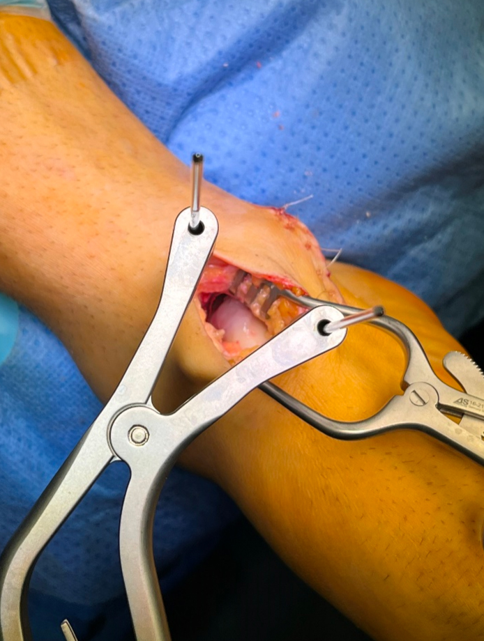
The surgical technique involved two phases; the first was arthroscopic and was needed for diagnostic confirmation and microfracture. Microfractures were performed systematically with a Chondro Pick (Arthrex, Naples, FL) with a depth of 4 mm and a distance of 3-4 mm between the perforations. The entire surface of the lesion was covered with perforations. The second phase included the arthrotomy, medial or lateral, based on the localization of the lesion (Figure 1), and the subsequent preparation and positioning of a bilayer collagen I/III membrane (Chondro-Gide, Geistlich Pharma AG, Wolhusen, Switzerland) (Figure 2), which was fixed with a synthetic fibrin glue (Figure 3). In two patients, due to the posterior location of the OLT, a medial malleolar osteotomy was required, which was subsequently fixed using two cannulated screws. Following these procedures, a drain was placed, layered suturing was performed, the surgical dressing was applied, and the limb was positioned in a 90° half-cast. The operation was generally carried out under spinal-general anesthesia with a tourniquet applied at the root of the limb. Ankle mobilization according to pain tolerance was allowed starting from the third postoperative day in order to recover the range of motion (ROM). The physiotherapy program aimed for full ROM recovery by the sixth postoperative week.
Postoperative radiographs of the ankle were obtained immediately after surgery, and at 2 and 6 weeks postoperatively. A follow-up MRI was performed between 6 and 10 weeks after the procedure.
Figure 1. Intraoperative view of the osteochondral lesion after arthrotomy.
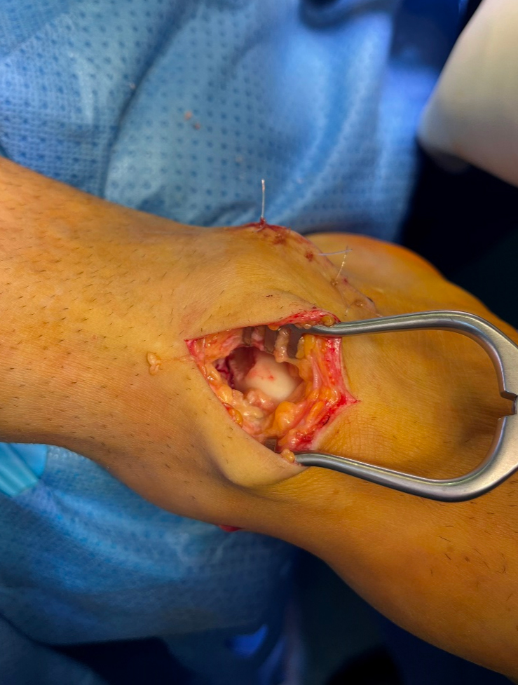
Figure 2. Positioning of the collagen membrane.
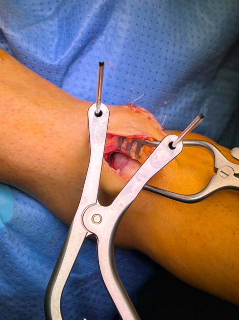
Figure 3. Fixation of the membrane with fibrin glue.
Outcome Measures
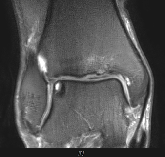
The clinical evaluation was performed by clinical examination and 3 patient-reported outcome measures (PROMs): the European Foot and Ankle Society (EFAS)18, Foot Function Index (FFI)19, and Manchester Oxford Foot Questionnaire (MOXFQ)20. All 3 were recorded at a mean of 10 years post-operatively. Radiologic follow-up was conducted using MRI and was performed with a 3T MRI unit. The MRI was performed post-operatively at 2 months follow-up (Figure 4), and approximately 10 years after surgery on average (Figure 5). In the final MRI, the Magnetic Resonance Observation of Cartilage Repair Tissue (MOCART) 2.021 score was evaluated independently by two radiologists specialized in musculoskeletal tissue analysis.
Figure 4. Preoperative MRI of the osteochondral lesion.
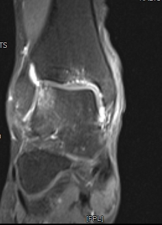
Figure 5. 10-year follow-up post-operative MRI of the lesion treated with AMIC.
Statistical Analysis
Descriptive statistics for the PROMs and the MOCART 2.0 parameters are presented as means and standard deviations. Inter-observer reliability between the two radiologists for the MOCART 2.0 score was assessed using Pearson’s correlation coefficient. To evaluate the relationship between the MOCART 2.0 score and the PROMs, both Pearson’s and Spearman’s rho correlation coefficients were calculated, given the small sample size. A p-value < 0.05 was considered statistically significant. The statistical analysis was conducted using JASP software version 0.19 (Amsterdam, Netherlands).
Ethics Approval
The Ethics Committee of the Review Board of Hessingpark Clinic approved the study protocol on January 31st, 2023 (Study Protocol: 01-INT-2023).
Results
The study included 19 patients (20 lesions) who had undergone AMIC for the treatment of OLT. The demographic data of the entire cohort are presented in Table 1.
Table 1. Demographic data of the patients.
| Gender | Female: 7 – Male: 12 |
| Age at surgery (years) | 47.3±10.4 |
| Lesion area (mm2) | 109.5±50.4 |
| BMI | 26.0±2.8 |
Imaging Results
Two radiologists separately examined the MRI performed 10 years after surgery and evaluated the MOCART 2.0 score. The values for the parameters of the MOCART 2.0 score are presented in Table 2. An analysis was therefore first conducted to evaluate the possible homogeneity of the values obtained by the observers. The Pearson correlation highlighted the significance of this correlation for most of the subcategories of the MOCART 2.0 score and, in particular, for the final result of the MOCART 2.0 score (Table 3). The categories filling, integration of adjacent cartilage, bone defect/overgrowth, subchondral changes, and total reported a significant correlation in the Pearson analysis.
Subsequently, for the correlation analysis, a MOCART 2.0 value was defined by averaging the observed values from each reviewer.
In addition to the MOCART 2.0 score, the radiologists separately assessed the presence of edema: in 3 patients, both radiologists reported the presence of this finding, while in 4 patients, this finding was assessed by only one radiologist. We therefore did not evaluate this parameter due to the discrepancy in the observations.
Table 2. Descriptive statistics of the Magnetic Resonance Observation of Cartilage Repair Tissue (MOCART) 2.0 score parameters, evaluated separately by two examiners.
| Reviewer 1 | Reviewer 2 | |
| Mean ± SD (range) | Mean ± SD (range) | |
| Volume of cartilage defect filling | 14±5.5 (0-20) | 13.5±7.9 (0-20) |
| Integration into adjacent cartilage | 12.8±3.8 (5-15) | 11.5±4.9 (0-15) |
| Surface of the repair tissue | 5.5±3.2 (0-10) | 5.5±3.6 (0-10) |
| Structure of the repair tissue | 0.5±2.2 (0-10) | 0.0±0.0 (0-0) |
| Signal intensity of the repair tissue | 4.0±5.0 (0-10) | 8.5±3.6 (0-10) |
| Bone defect/overgrowth | 2.5±2.6 (0-5) | 3.8±2.2 (0-5) |
| Subchondral changes | 3.3±6.1 (0-20) | 3.8±6.3 (0-20) |
| Total | 46.5±13.3 (20-60) | 44.8±12.1 (20-65) |
Table 3. Pearson’s correlation of the Magnetic Resonance Observation of Cartilage Repair Tissue (MOCART) 2.0 score analyzed by two examiners.
| MOCART 2.0 parameters | Pearson’s r | p | Sig. |
| Volume of cartilage defect filling | 0.622 | 0.003 | * |
| Integration into adjacent cartilage | 0.475 | 0.034 | * |
| Surface of the repair tissue | 0.320 | 0.169 | |
| Structure of the repair tissue | – | – | * The variance was equal to 0 |
| Signal intensity of the repair tissue | 0.343 | 0.139 | |
| Bone defect/overgrowth | 0.577 | 0.008 | * |
| Subchondral changes | 0.935 | <0.001 | * |
| Total | 0.616 | 0.004 | * |
Clinical Results
With regard to the clinical outcomes, 3 PROMs were administered to the patients to evaluate their return to normal daily activities. These are summarized in Table 4.
Table 4. Descriptive statistics of PROMs results.
| Mean ± SD (range) | |
| EFAS | 19.8±4.6 (9-24) |
| EFAS sport | 12.1±4.9 (1-16) |
| EFAS total | 31.9±9.1 (12-40) |
| MOXFQ walking standing | 4.5±6.4 (0-21) |
| MOXFQ pain | 3.2±3.8 (0-10) |
| MOXFQ social interaction | 1.9±3.3 (0-11) |
| MOXFQ total | 9.6±12.9 (0-37) |
| FFI pain | 5.9±8.2 (0-23) |
| FFI function | 10.3±13.6 (0-39) |
| FFI total | 16.2±20.9 (0-59) |
European Foot and Ankle Society (EFAS), Manchester Oxford Foot Questionnaire (MOXFQ), Foot Function Index (FFI).
Correlations
The relationship between the MOCART 2.0 score and the various PROMs was assessed, and these are presented in Table 5. While the outcome scores as well as MOCART are parametric data, which would typically call for a Pearson’s correlation, with the small sample size, we also calculated a Spearman-rho correlation coefficient. As shown in Table 5, there was no relationship between the MOCART scores and any of the PROMs.
Revisions and Reoperations
Among the 19 patients of the study, no revision or major or minor complication was reported at the follow-up.
Table 5. Correlations between the Magnetic Resonance Observation of Cartilage Repair Tissue (MOCART) 2.0 and the PROMs.
| Pearson | Spearman | |||
| r | p | r | p | |
| MOCART 2.0 – EFAS | 0.283 | 0.226 | 0.226 | 0.339 |
| MOCART 2.0 – FFI | 0.329 | 0.157 | 0.325 | 0.163 |
| MOCART 2.0 – MOXFQ | -0.302 | 0.195 | -0.233 | 0.323 |
European Foot and Ankle Society (EFAS), Manchester Oxford Foot Questionnaire (MOXFQ), Foot Function Index (FFI), Magnetic Resonance Observation of Cartilage Repair Tissue (MOCART).
Discussion
Clinical Outcomes
The results of all 3 PROMs indicate that patients treated with AMIC for OLT showed good clinical results up to 10 years post-operatively. The scores for MOXFQ, EFAS and FFI, along with their subscales, demonstrate that patients experienced durable, positive outcomes with regard to both pain and function. While the radiological evaluations yielded a fairly low score, no correlation was found between any of the PROMs and the MOCART scores.
The outcome scores in this study were comparable to what has been reported elsewhere for surgical repair of OLT. Although the EFAS is a relatively recent assessment tool, its relation to well-established outcome scores has already been demonstrated22. Furthermore, the scores we have documented in this study are apparently slightly better than those published22-24. The EFAS scores, along with the specific subscales, that we presented show slightly better results compared with published data, with a similar comparison of our FFI results23,24, while the MOXFQ outcomes that we have measured are notably lower (indicating a better outcome) than those reported in the literature22. The sample size in this study is small, but the length of follow-up in our data supports the durability of the AMIC technique for the surgical repair of chondral defects.
The utility of this surgical technique in the repair of OLT has been reported previously, in various surgical approaches25,26. Regardless of the specific surgical techniques, positive outcomes have been reported in studies that have documented medium-term follow-up of 5 years23,27,28. In terms of patients treated, these positive results have been reported in a study that included a large number of patients (n=129) with a 5-year follow-up23. Additionally, a study29 with 8 years of follow-up concluded that the AMIC procedure led to a significant reduction of pain, a recovery of ankle function, and a successful return to sports. While not an exhaustive list of data concerning AMIC, the length of follow-up and the number of successfully treated patients support the use of this technique, and the data we have presented further underscore the procedure’s potential to provide lasting, positive outcomes for patients.
Radiological Evaluations
Regarding MRI evaluations, the utility of this imaging modality for evaluating outcomes has already been questioned. It has been succinctly stated that clinical outcome does not correlate with any MRI parameter, even though the patients had positive outcomes as measured by the American Orthopedic Foot & Ankle Society (AOFAS)30. To further question the utility of this imaging modality, it was noted that there was no relationship between the MOCART score and any of the PROMs31, which was also a conclusion of a study that presented 8 years of follow-up after OLT repair via the AMIC procedure29. As our data show, there is no correlation between the PROMs and the MOCART 2.0. This should be kept in mind, especially by colleagues who do not frequently deal with the repair of OLT. An MRI performed for simple follow-up may show a MOCART/MOCART 2.0 score that is concerning enough to warrant another surgery. However, the clinical and anamnestic evaluation of the subject is much more important, as it can indicate the success or failure of the primary surgical intervention. The importance of MRI in post-operative routine check-ups should thus be reconsidered, as it does not seem to provide any real benefit to patients or physicians.
Limitations
It is important to acknowledge the limitations of this study. Firstly, the small sample size precluded a statistical analysis that could have considered covariates such as age, body mass index (BMI), or defect size and whether these variables had an impact on the measured outcomes. Another limitation is the lack of preoperative data, as this would have given a precise value of the clinical improvement of the patients, and allowed an assessment of the positive response rate in comparison to established values for minimal clinically important difference.
Conclusions
The observational results that have been presented in this study, both clinical and radiological, are comparable with other AMIC studies in the literature, and this data extends the documented follow-up to 10 years22-24. The data presented, although with the limitations mentioned above, show good long-term results for the AMIC technique. Larger datasets and multicenter studies are needed to provide more robust evidence supporting the conclusions of this study.
Ethics Approval
This study was conducted in accordance with the Declaration of Helsinki of 1975 (as revised in 2013), and the protocol was reviewed and approved by the Review Board of Hessingpark Clinic (Study Protocol: 01-INT-2023, date of approval: January 31st, 2023).
Informed Consent
All subjects provided written informed consent for inclusion before they participated in the study.
Acknowledgments
A sincere acknowledgment to the Hessingpark Clinic staff who helped the authors during the patient follow-up.
Funding
This research did not receive any specific grant from funding agencies in the public, commercial, or not-for-profit sectors.
Authors’ Contributions
E.D. and M.T. participated in the drafting and critical revision of the manuscript. M.J. contributed extensively to the study’s methodology development and was instrumental in the acquisition of data. W.F. and S.H. played a key role in obtaining the radiological data and their interpretation. E.D. provided substantial input in the writing and editing of the manuscript and was also deeply involved in the data analysis and interpretation. M.T. contributed significantly to the drafting of the manuscript, particularly in the discussion and conclusion sections.
ORCID ID
Elena Delmastro: 0000-0003-1710-8635
Conflict of Interest
The authors declare that they have no conflict of interest to disclose.
Data Availability
The datasets generated during and/or analyzed during the current study are available from the corresponding author upon reasonable request.
AI Disclosure
Artificial intelligence or assisted technologies were not used in the preparation of this study.
References
- Cha SD, Kim HS, Chung ST, Yoo JH, Park JH, Kim JH, Hyung JW. Intra-articular lesions in chronic lateral ankle instability: comparison of arthroscopy with magnetic resonance imaging findings. Clin Orthop Surg 2012; 4: 293-299.
- Murawski CD, Kennedy JG. Operative treatment of osteochondral lesions of the talus. J Bone Joint Surg Am 2013; 95: 1045-1054.
- Kerkhoffs GMMJ, Karlsson J. Osteochondral lesions of the talus. Knee Surg Sports Traumatol Arthrosc 2019; 27: 2719-2720.
- Lauck BJ, Shaffrey I, Anastasio AT, O’Neill CN, Hanselman AE, Adams SB. Twelve-Year Progression of Osteochondral Lesions of the Talus Observed Using Magnetic Resonance Imaging. Diagnostics (Basel) 2024; 14: 1251.
- Dahmen J, Karlsson J, Stufkens SAS, Kerkhoffs GMMJ. The ankle cartilage cascade: incremental cartilage damage in the ankle joint. Knee Surg Sports Traumatol Arthrosc 2021; 29: 3503-3507.
- Kim J, Cho H, Young K, Park J, Lee J, Suh D. In vivo animal study and clinical outcomes of autologous atelocollagen-induced chondrogenesis for osteochondral lesion treatment. J Orthop Surg Res 2015; 10: 82.
- Duramaz A, Baca E. Microfracture provides better clinical results than debridement in the treatment of acute talar osteochondral lesions using arthroscopic assisted fixation of acute ankle fractures. Knee Surg Sports Traumatol Arthrosc 2018; 26: 3089-3095.
- Migliorini F, Eschweiler J, Maffulli N, Schenker H, Driessen A, Rath B, Tingart M. Autologous Matrix Induced Chondrogenesis (AMIC) Compared to Microfractures for Chondral Defects of the Talar Shoulder: A Five-Year Follow-Up Prospective Cohort Study. Life (Basel) 2021; 11: 244.
- Lee KT, Song SY, Hyuk J, Kim SJ. Lesion Size May Predict Return to Play in Young Elite Athletes Undergoing Microfracture for Osteochondral Lesions of the Talus. Arthroscopy 2021; 37: 1612-1619.
- Steele JR, Dekker TJ, Federer AE, Liles JL, Adams SB, Easley ME. Republication of “Osteochondral Lesions of the Talus: Current Concepts in Diagnosis and Treatment.” Foot Ankle Orthop 2023; 8: 24730114231192961.
- Feeney KM. The Effectiveness of Osteochondral Autograft Transfer in the Management of Osteochondral Lesions of the Talus: A Systematic Review and Meta-Analysis. Cureus 2022; 14: e31337.
- Lenz CG, Tan S, Carey AL, Ang K, Schneider T. Matrix-Induced Autologous Chondrocyte Implantation (MACI) Grafting for Osteochondral Lesions of the Talus. Foot Ankle Int 2020; 41: 1099-1105.
- Migliorini F, Maffulli N, Bell A, Hildebrand F, Weber CD, Lichte P. Autologous Matrix-Induced Chondrogenesis (AMIC) for Osteochondral Defects of the Talus: A Systematic Review. Life (Basel) 2022; 12: 1738.
- Walther M, Gottschalk O, Aurich M. Operative management of osteochondral lesions of the talus: 2024 recommendations of the working group ‘clinical tissue regeneration’ of the German Society of Orthopedics and Traumatology (DGOU). EFORT Open Rev 2024; 9: 217-234.
- Walther M, Valderrabano V, Wiewiorski M, Usuelli FG, Richter M, Baumfeld TS, Kubosch J, Gottschalk O, Wittmann U. Is there clinical evidence to support autologous matrix-induced chondrogenesis (AMIC) for chondral defects in the talus? A systematic review and meta-analysis. Foot Ankle Surg 2021; 27: 236-245.
- Migliorini F, Maffulli N, Schenker H, Eschweiler J, Driessen A, Knobe M, Tingart M, Baroncini A. Surgical Management of Focal Chondral Defects of the Talus: A Bayesian Network Meta-analysis. Am J Sports Med 2022; 50: 2853-2859.
- Albert BL, Harty M. Transchondral fractures (osteochondritis dissecans) of the talus. J Bone Joint Surg Am 1959; 41-A: 988-1020.
- Richter M, Agren PH, Besse JL, Cöster M, Kofoed H, Maffulli N, Rosenbaum D, Steultjens M, Alvarez F, Boszczyk A, Buedts K, Guelfi M, Liszka H, Louwerens JW, Repo JP, Samaila E, Stephens M, Witteveen AGH. EFAS Score – Multilingual development and validation of a patient-reported outcome measure (PROM) by the score committee of the European Foot and Ankle Society (EFAS). Foot Ankle Surg 2018; 24: 185-204.
- Naal FD, Impellizzeri FM, Huber M, Rippstein PF. Cross-Cultural Adaptation and Validation of the Foot Function Index for Use in German-Speaking Patients with Foot Complaints. Foot Ankle Int 2008; 29: 1222-1231
- Dawson J, Coffey J, Doll H, Lavis G, Cooke P, Herron M, Jenkinson C. A patient-based questionnaire to assess outcomes of foot surgery: validation in the context of surgery for hallux valgus. Qual Life Res 2006; 15: 1211-1222.
- Schreiner MM, Raudner M, Marlovits S, Bohndorf K, Weber M, Zalaudek M, Röhrich S, Szomolanyi P, Filardo G, Windhager R, Trattnig S. The MOCART (Magnetic Resonance Observation of Cartilage Repair Tissue) 2.0 Knee Score and Atlas. Cartilage 2021; 13: 571S-587S.
- Gottschalk O, Kerschl FW, Mazet J, Suero E, Hörterer H, Altenberger S, Böcker W, Walther M. Evaluation of the new defined EFAS score (European foot and ankle society score) in relation to already established functional scores (FFI, MOXFQ) after cartilage reconstructive therapy (AMIC®) for osteochondral lesion of the talus. Foot Ankle Surg 2022; 28: 975-978.
- Richter M, Zech S, Meissner S, Naef I. Autologous matrix induced chondrogenesis plus peripheral blood concentrate (AMIC+PBC) in chondral lesions at the ankle as part of a complex surgical approach – 5-year follow-up. Foot Ankle Surg 2022; 28: 1321-1326.
- Deiss L, Walther M, Pfahl K, Hörterer H, Mehlhorn A, Röser A, Gottschalk O. Long-Term Results after Autologous Matrix-Induced Chondrogenesis for Osteochondral Lesions of the Talus: A 10-Year Cohort Study. Cartilage 2024; 12: 19476035241301896.
- Galla M, Duensing I, Kahn TL, Barg A. Open reconstruction with autologous spongiosa grafts and matrix-induced chondrogenesis for osteochondral lesions of the talus can be performed without medial malleolar osteotomy. Knee Surg Sports Traumatol Arthrosc 2019; 27: 2789-2795.
- Sadlik B, Kolodziej L, Puszkarz M, Laprus H, Mojzesz M, Whyte GP. Surgical repair of osteochondral lesions of the talus using biologic inlay osteochondral reconstruction: Clinical outcomes after treatment using a medial malleolar osteotomy approach compared to an arthroscopically-assisted approach. Foot Ankle Surg 2019; 25: 449-456.
- Gottschalk O, Altenberger S, Baumbach S, Kriegelstein S, Dreyer F, Mehlhorn A, Hörterer H, Töpfer A, Röser A, Walther M. Functional Medium-Term Results After Autologous Matrix-Induced Chondrogenesis for Osteochondral Lesions of the Talus: A 5-Year Prospective Cohort Study. J Foot Ankle Surg 2017; 56: 930-936.
- Götze C, Nieder C, Felder H, Peterlein CD, Migliorini F. AMIC for traumatic focal osteochondral defect of the talar shoulder: a 5 years follow-up prospective cohort study. BMC Musculoskelet Disord 2021; 22: 638.
- Weigelt L, Hartmann R, Pfirrmann C, Espinosa N, Wirth SH. Autologous Matrix-Induced Chondrogenesis for Osteochondral Lesions of the Talus: A Clinical and Radiological 2- to 8-Year Follow-up Study. Am J Sports Med 2019; 47: 1679-1686.
- Casari FA, Germann C, Weigelt L, Wirth S, Viehöfer A, Ackermann J. The Role of Magnetic Resonance Imaging in Autologous Matrix-Induced Chondrogenesis for Osteochondral Lesions of the Talus: Analyzing MOCART 1 and 2.0. Cartilage 2021; 13(1_suppl): 639S-645S.
- Gottschalk O, Mazet J, Kerschl F, Schenk H, Suero EM, Hörterer H, Böcker W, Walther M. Correlation between EFAS- and MOCART score and clinical outcome after AMIC®-procedure for osteochondral lesion of the talus. Arch Orthop Trauma Surg 2023; 143: 2895-2900.
To cite this article
Osteochondral lesions of the talus treated with Autologous Matrix-Induced Chondrogenesis: long-term results
JOINTS 2026;
4: e2154
DOI: 10.26355/joints_202607_2154
Publication History
Submission date: 08 Oct 2025
Revised on: 30 Oct 2025
Accepted on: 16 Jun 2026
Published online: 30 Jul 2026