JOINTS 2026;
4: e2191
DOI: 10.26355/joints_202610_2191
Double-level femoral and tibial derotational osteotomy for recurrent patellar instability: a systematic review
Topic: Osteotomy
Category: Systematic Review
Abstract
OBJECTIVE: Recurrent patellar instability complicated by combined femoral and tibial torsional deformities presents a complex therapeutic challenge. Although double-level derotational osteotomy has been proposed as an effective surgical option, evidence remains limited. This systematic review aimed to evaluate clinical, radiological, and functional outcomes of combined femoral and tibial derotational osteotomies in patients with recurrent patellar instability.
MATERIALS AND METHODS: A systematic search of PubMed, Scopus, Embase, MEDLINE and Cochrane was performed according to PRISMA 2020 guidelines. Studies published from 1996 to April 2025 reporting double-level derotational osteotomy for patellar instability, with a minimum individual follow-up of at least 24 months and Level of Evidence I-IV, were included. Data extraction included demographic characteristics, radiographic torsional parameters, clinical outcome scores, recurrent patellar instability, complications, and fixation methods. Demographic variables were summarized using weighted means when appropriate, whereas clinical and radiographic outcomes were summarized descriptively due to methodological heterogeneity across studies. Risk of bias was assessed using the Risk Of Bias In Non-randomized Studies of Interventions (ROBINS-I) tool, while complications were graded according to the Clavien–Dindo classification.
RESULTS: Seven retrospective studies comprising 64 patients (79 limbs) were included. The weighted mean patient age was 19.3 years, with a weighted mean follow-up of 45.2 months. Femoral and tibial rotational corrections demonstrated substantial improvements in rotational alignment. Significant radiographic improvements were also reported for patellar tilt and torsional parameters. Clinical and functional outcomes generally improved postoperatively; however, in some studies including both single- and double-level procedures, subgroup-specific outcomes were unavailable and were reported only for the overall mixed cohort. Complication rates were low, with isolated cases of nonunion, fixation failure, postoperative stiffness, and one recurrent patellar dislocation reported at long-term follow-up.
CONCLUSIONS: Double-level derotational osteotomy appears to be a safe and effective option for carefully selected patients with recurrent patellar instability and severe combined torsional deformities. However, current evidence is limited by methodological heterogeneity and small retrospective cohorts, highlighting the need for standardized diagnostic criteria and prospective multicenter studies.
Introduction
Recurrent patellar instability is a complex, multifactorial condition that predominantly affects adolescents and young adults, especially females, with recurrence rates up to 50% after an initial dislocation in the presence of anatomical risk factors such as trochlear dysplasia, patella alta, and elevated tibial tubercle–trochlear groove (TT–TG) distance1. Additionally, increased femoral anteversion and external tibial torsion have been identified as significant contributors to abnormal patellar tracking and recurrent instability2,3.
Soft-tissue reconstructions, such as medial patellofemoral ligament (MPFL) repair, may be insufficient when torsional deformities exceed 20-25°, resulting in suboptimal outcomes2,4. Advanced preoperative imaging, including low-dose computed tomography (CT) and EOS systems, has improved diagnostic accuracy and enabled more precise surgical planning5.
Derotational osteotomies at the femoral and tibial levels have emerged as important interventions. Derotational distal femoral osteotomy (D-DFO), with or without concomitant procedures, has shown significant improvements in pain and functional scores in patients with femoral anteversion >20°6,7. Recent evidence suggests that combined femoral and tibial derotational osteotomies provide superior functional outcomes and greater reduction in anterior knee pain compared with tibial tubercle transfer (TTT) combined with medial patellofemoral ligament (MPFL) reconstruction in patients with severe torsional deformities1-3.
Nevertheless, evidence remains limited; most available studies are retrospective case series or cohort trials. Key parameters, such as optimal correction thresholds, complication rates, and long-term outcomes, have not been definitively established8,9. Furthermore, technical complexity and potential morbidity demand improved selection criteria and operative consistency.
Despite the increasing use of double-level derotational osteotomy, the available evidence remains fragmented, consisting predominantly of small retrospective case series with heterogeneous patient selection, surgical indications, radiological assessment methods, and outcome reporting. To date, no systematic review has specifically synthesized the evidence regarding the clinical, radiological, and functional outcomes of this procedure in patients with recurrent patellar instability.
This systematic review aims to summarize current clinical, radiologic, and functional outcomes of double-level femoral and tibial derotational osteotomies in recurrent patellar instability, to define clear surgical indications, evaluate effectiveness, and identify areas for future research.
Materials and Methods
Research Question
This systematic review was conducted in accordance with the 2020 PRISMA guidelines to identify and select relevant studies10. To minimize bias, two independent reviewers (RGV and FB) conducted the search and selection, and uncertainties were resolved by consulting a third author (MC).
Inclusion and Exclusion Criteria
Eligible studies concerned patients undergoing double-level osteotomy for patellar instability. To be considered eligible, studies had to involve human subjects, be published between 1996 and April 2025, report a minimum follow-up of at least 24 months for the included patients, and provide a level of evidence (LoE) between I and IV. Studies were excluded if they were biochemical or in vitro investigations, case reports, editorials, book chapters, technical notes, preclinical studies, or Level V evidence, in order to include only studies providing clinically relevant evidence.
Search Strategy and Study Screening
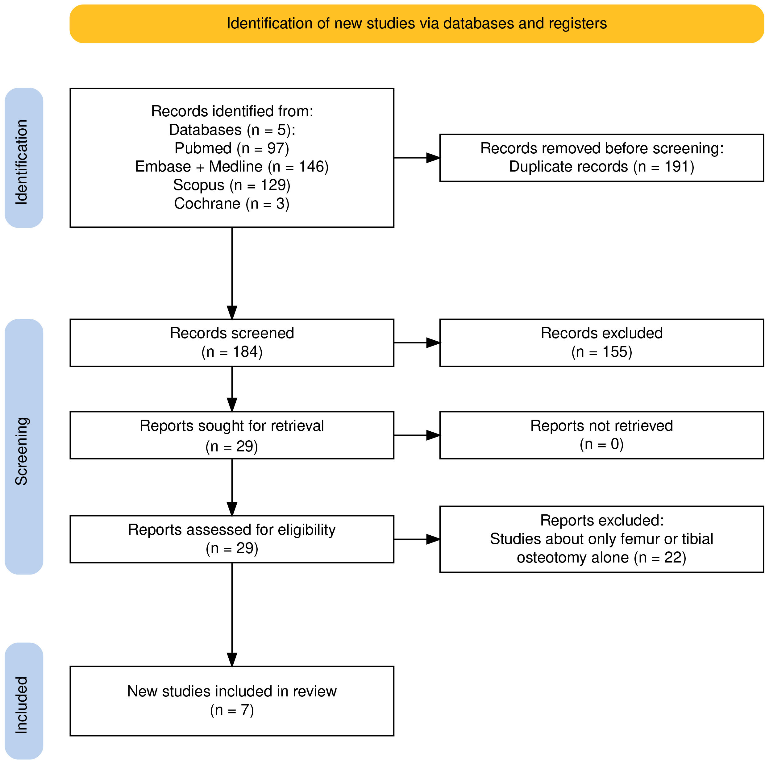
A thorough search of five large databases (PubMed, Scopus, Embase, MEDLINE and Cochrane) was carried out using the following MeSH terms: ((patellar instability) OR (recurrent patellar dislocation) OR (patellar dislocation) OR (patellofemoral instability)) AND ((derotational osteotomy) OR (torsional osteotomy) OR (femoral derotation) OR (tibial derotation) OR (derotation osteotomy)) AND ((outcome*) OR (result*) OR (surgical treatment) OR (surgical technique)). This search generated 375 articles. After deleting duplicates, 184 articles remained. Screening of titles and abstracts led to the exclusion of 155 studies. Full-text reviews were conducted for the remaining 29 articles; ultimately, seven studies3,11-16 met the inclusion criteria for the qualitative analysis. These selected studies provided epidemiological data, radiographic data on femoral and tibial torsion before and after surgery, functional outcomes, range of motion, and complication rates. The selection process is illustrated in the PRISMA flow chart (Figure 1).
Assessment of Methodological Quality
The level of evidence of each study was classified according to the Oxford Center for Evidence-Based Medicine 2011 framework17. Risk of bias was independently assessed by two reviewers using the Risk Of Bias In Non-randomized Studies of Interventions (ROBINS-I) tool, which evaluates seven domains of bias: confounding, selection of participants, classification of interventions, deviations from intended interventions, missing data, measurement of outcomes, and selection of the reported result18. Disagreements were resolved through discussion with a third reviewer. Complications were classified according to the Clavien–Dindo system19. This systematic review was prospectively registered in the International Prospective Register of Systematic Reviews (PROSPERO; registration number: CRD420251038762)20.
Data Extraction
The data extracted from the included articles were systematically recorded using a standardized data extraction template, which included the following variables: author and year of publication, study design, patient sample size, average age of participants, body mass index (when available), duration of follow-up, surgical site of correction, pre- and postoperative radiographic measurements, pre- and postoperative clinical outcome scores, complications, and details of the fixation system used for osteotomy stabilization. For studies reporting both single-level and double-level osteotomies, data from patients who underwent double-level derotational osteotomy were extracted whenever these data were reported separately. When subgroup-specific clinical outcomes were unavailable, outcomes reported for the overall mixed cohort were identified as such and were not considered specifically attributable to double-level osteotomy.
Data Analysis
Data analyses were performed using R software (2022, version 4.1.3; R Core Team, Vienna, Austria). Descriptive statistics were used to analyze the collected data. When appropriate, weighted means for demographic variables were calculated using the number of patients included in each study as the weighting factor. Continuous variables were summarized as weighted means whenever sufficient study-level data were available; otherwise, they were reported descriptively using means and ranges as provided in the original studies. Standard deviations (SDs) were reported only when available in the original publications. Absolute numbers and frequency distributions were calculated for categorical data.
Results
Study Characteristics
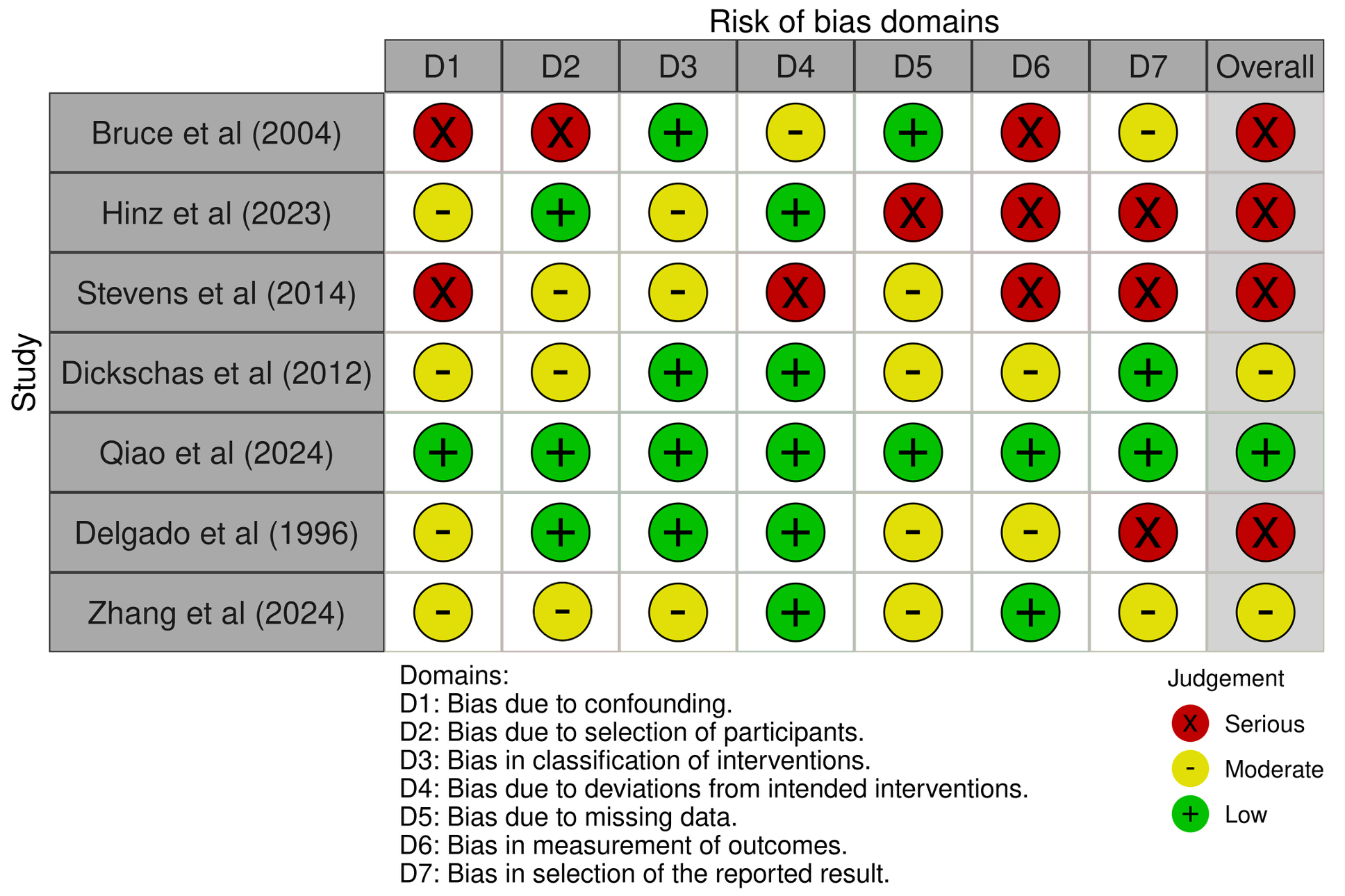
In line with PRISMA guidelines, seven retrospective studies3,11-16 were included in the final analysis, comprising 64 patients and 79 operated limbs. The cohorts were predominantly female, with over 60% female representation. The weighted mean patient age across the included studies was 19.3 years, while the weighted mean follow-up was 45.2 months. Detailed demographic characteristics are summarized in Table 1. Risk-of-bias assessment using the ROBINS-I tool demonstrated variable methodological quality across the included studies. One study3 was judged to have an overall low risk of bias, two studies11,13 showed a moderate risk of bias, and four studies12,14-16 were considered at serious risk of bias, mainly because of confounding, participant selection, outcome measurement, and selective reporting (Figure 2). Reported complications were predominantly low grade according to the Clavien–Dindo classification19, with most events managed without long-term sequelae.
Table 1. Demographic and clinical characteristics of the included studies.
| Author | Year | Study design | Number of patients | Number of limbs | Laterality | Laterality (R/L/Bil) | Gender | Age | BMI, kg/m2
|
Follow-up, months | |||
| Right (n/%) | Left (n/%) | Bilateral (n/%) | Male (n/%) | Female (n/%) | Mean (Range) | Mean (Range) | Mean (Range) | ||||||
| Bruce and Stevens16 | 2004 | Retrospective | 14 | 27 | NA | NA | 13 (92.9%) | — / — / 13 | 1 (7.1%) | 13 (92.9%) | 14.9 (11.75-18) | NA | 62.4 (24-144) |
| Hinz et al14 | 2023 | Retrospective | 2 | 2 | NA | NA | 0 (0%) | — / — / 0 | NA | NA | 23.5 (19.8-29.0) | 24.3 (19.6-29.0) | 38 (31.8-52.5) |
| Stevens et al15 | 2014 | Retrospective | 8 | 8 | 4 (50.0%) | 4 (50%) | 0 (0%) | 4 / 4 / 0 | 3 (37.5%) | 5 (62.5%) | 17.5 (9-24) | NA | 55.1 (17-113) |
| Dickschas et al11 | 2012 | Retrospective | 2 | 2 | 2 (100.0%) | 0 (0%) | 0 (0%) | 2 / 0 / 0 | 1 (50.0%) | 1 (50.0%) | 19.5 (15-24) | 24.9 | NA |
| Qiao et al3 | 2024 | Retrospective | 18 | 18 | 7 (38.9%) | 11 (61.1%) | 0 (0%) | 7 / 11 / 0 | 3 (16.7%) | 15 (83.3%) | 20.7 (16-30) | NA | 29 (17.9-31.6) |
| Delgado et al12 | 1996 | Retrospective | 5 | 7 | 2 (28.6%) | 1 (14.3%) | 2 (40%) | 2 / 1 / 2 | 3 (60.0%) | 2 (40.0%) | 14.4 (11-18) | NA | 33.6 (24-48) |
| Zhang et al13 | 2024 | Retrospective | 15 | 15 | NA | NA | 0 (0%) | — / — / 0 | 1 (6.7%) | 14 (93.3%) | 23.8 (17.2-30.4) | NA | 48 (27.6-68.4) |
R = right; L = left; Bil = bilateral; NA = not available; n/% = number (percentage); BMI = body mass index.
Figure 2. Risk-of-bias assessment of the included studies according to the ROBINS-I tool.
Surgical Indications and Techniques
The surgical site of correction varied among the included studies and involved distal femoral, proximal tibial, supramalleolar tibial, or combined femoral and tibial derotational osteotomies, depending on the underlying deformity and surgical indication. Across the included studies, the indication for double-level derotational osteotomy was recurrent patellar instability associated with combined femoral and tibial torsional deformities. Although the threshold values varied among studies, surgical indication was generally based on the presence of clinically relevant combined femoral and tibial torsional deformities, supported by comprehensive clinical evaluation and CT-based assessment of rotational alignment. The primary objective of double-level derotational osteotomy was to restore physiological lower-limb rotational alignment, improve patellar tracking and stability, and reduce anterior knee pain. Femoral correction was most commonly performed at the distal femur, whereas tibial correction was carried out at either the proximal tibia or the supramalleolar level according to the underlying deformity.
Radiographic Outcomes
Radiographic parameters were reported heterogeneously across the included studies. Radiological assessment of torsional deformities was also heterogeneous. Most studies used computed tomography (CT) to quantify femoral anteversion and external tibial torsion; however, different measurement protocols and anatomical reference axes were adopted across studies, limiting direct comparison of absolute rotational values. Femoral and tibial rotational correction angles were available in four studies3,11-13 and demonstrated substantial correction following surgery (Table 2). In Dickschas et al11, only two patients underwent bifocal osteotomy. The reported femoral corrections were 12° and 23°, while the corresponding tibial corrections were 10° and 12°, respectively. Patellar tilt angles were reported in two studies3,13, both of which demonstrated significant postoperative correction. Qiao et al3 observed a reduction from 37.1° (32.7-41.6) preoperatively to 2.3° (-1.7 to 6.2) postoperatively, while Zhang et al13 reported a decrease from 37° (9-65) to 11° (1-21), corresponding to a mean correction of 26° to 34.8°.
Table 2. Radiographic measurements of femoral torsion, tibial torsion and patellar tilt before and after double-level derotational osteotomy.
| Author | Year | Femoral Torsion, ° | Tibial Torsion, ° | Patellar tilt angle, ° | ||||||
| Preoperative | Postoperative | Correction, ° | Preoperative | Postoperative | Correction, ° | Preoperative | Postoperative | Correction, ° | ||
| Mean (range) | Mean (range) | Mean (range) | Mean (range) | Mean (range) | Mean (range) | |||||
| Bruce and Stevens16 | 2004 | 29 (20-40) | 27 (20-35) | |||||||
| Hinz et al14 | 2023 | |||||||||
| Stevens et al15 | 2014 | 25 (30-35) | 29.4 (20-40) | |||||||
| Dickschas et al11* | 2012 | 12, 23 | 10, 12 | |||||||
| Qiao et al3 | 2024 | 31.9 (30.8 to 32.9) | 8.6 (6.3 to 11) | 23.3 | 34.2 (32.7-35.6) | 9.6 (7.4-11.9) | 24.6 | 37.1 (32.7-41.6) | 2.3 (-1.7 to 6.2) | 34.8 |
| Delgado et al12 | 1996 | 35 | 22 | |||||||
| Zhang et al13 | 2024 | 37.2 | 5.3 | 31.9 | 37 (9 to 65) | 11 (1 to 21) | 26 | |||
° = degree; * = Only the rotational corrections specifically reported for the two patients undergoing bifocal osteotomy are presented (femoral correction: 12° and 23°; tibial correction: 10° and 12°); subgroup-specific pre and postoperative torsional values were not available.
Clinical Outcomes
Clinical outcomes were reported heterogeneously across the included studies (Table 3). Functional outcomes were assessed using Kujala, Lysholm, International Knee Documentation Committee (IKDC), Tegner, Knee injury and Osteoarthritis Outcome Score (KOOS), and Visual Analog Scale (VAS) scores, with postoperative improvements generally reported at short- to mid-term follow-up. However, in studies including both single- and double-level osteotomies, subgroup-specific clinical outcomes were not always available. Dickschas et al11 and Stevens et al15 reported clinical outcomes for the overall mixed cohort rather than separately for patients undergoing double-level osteotomy; therefore, these findings cannot be specifically attributed to the double-level procedure. Pain outcomes generally improved in the studies reporting VAS scores. Hinz et al14 observed a reduction from a median preoperative VAS of 2 (1-5) to 0 (0-1), while Qiao et al3 reported a reduction from 4.8 (1.8-7.8) to 1.4 (-0.6 to 3.4). Stevens et al15 and Dickschas et al11 reported decreases from 8.6 to 3.3 and from 7.3 to 2.6, respectively; however, these values were derived from their overall mixed cohorts. Kujala scores were available in three studies3,13,14 and improved across all cases. Hinz et al14 noted a postoperative increase from 55.6 (28.4-82.8) to 80.3 (46.9-113.7), and Qiao et al3 reported an increase from 64 (33.4-94.6) to 86.8 (68.6-105). Zhang et al13 and Delgado et al12 also reported postoperative scores ≥80. Lysholm and IKDC scores followed a similar trend, with Qiao et al3 demonstrating improvements in Lysholm (from 62 to 84.3) and IKDC (from 59.2 to 82.2), while Zhang et al13 and Delgado et al12 reported final scores exceeding 84. Tegner activity scores were reported in four studies3,11,13,14; however, the Tegner data reported by Dickschas et al11 referred to the overall mixed cohort and were not available separately for the double-level subgroup.
Complications
Overall, only one recurrent patellar dislocation was reported across all included studies. Complications were uncommon and were generally minor or successfully managed without long-term sequelae. Three studies11,12,14 reported no adverse events. Bruce and Stevens16 documented the highest complication rate (11.1%), including one case each of tibial fixation loss, femoral fracture, and fibular nonunion. Stevens et al15 and Qiao et al3 each reported one femoral nonunion (12.5% and 5.6%, respectively). Zhang et al13 reported one case of postoperative knee stiffness (6.7%) that resolved following rehabilitation. A comprehensive summary of clinical outcomes and complications is presented in Table 3.
Table 3. Reported functional outcomes and complications in studies including double-level derotational osteotomy for recurrent patellar instability.
| Author | Year | VAS score | Kujala score | Lysholm score | IKDC score | Tegner score | Complications | |||||
| Preoperative | Postoperative | Preoperative | Postoperative | Preoperative | Postoperative | Preoperative | Postoperative | Preoperative | Postoperative | |||
| Mean (range) | Mean (range) | Mean (range) | Mean (range) | Mean (range) | Mean (range) | Mean (range) | Mean (range) | Mean (range) | Mean (range) | N (%) | ||
| Bruce and Stevens16 | 2004 | 3 (11.1%) | ||||||||||
| Hinz et al14* | 2023 | 2 (1-5) | 0 (0-1) | 55.6 (28.4-82.8) | 80.3 (46.9-113.7) | 58.6 (23.8-93.4) | 79.5 (46.3-112.7) | 54.6 (17.2-92) | 74.1 (44.1-104.1) | 3 (3-4) | 4 (3-5) | 0 (0%) |
| Stevens et al15* | 2014 | 8.6 (7.9-9.4) | 3.3 (2-4.6) | 1 (12.5%) | ||||||||
| Dickschas et al11* | 2012 | 7.3 | 2.6 | 56.7 | 83.7 | 3.6 | 4.4 | 0 (0%) | ||||
| Qiao et al3 | 2024 | 4.8 (1.8-7.8) | 1.4 (-0.6-3.4) | 64 (33.4-94.6) | 86.8 (68.6-105) | 62 (31.8-92.2) | 84.3 (62.3-106.3) | 59.2 (33.8-84.6) | 82.2 (69.4-95) | 2.3 (0.7-3.9) | 4.1 (2.1-6.1) | 1 (5.6%) |
| Delgado et al12 | 1996 | 60 | 82 | 66 | 84 | 0 (0%) | ||||||
| Zhang et al13 | 2024 | 60 (42-78) | 82 (62-102) | 66 (40-92) | 84 (56-112) | 3 (1-5) | 3 (2-6) | 1 (6.7%) | ||||
VAS = Visual Analog Scale; IKDC = International Knee Documentation Committee; n/% = number (percentage); * = Clinical outcome scores were reported for the overall mixed cohort including both single- and double-level osteotomies and were not available separately for the double-level subgroup.
Discussion
The principal finding of this systematic review is that, although double-level derotational osteotomy appears to improve radiographic alignment and clinical outcomes in carefully selected patients, the currently available evidence is characterized by substantial methodological heterogeneity that limits definitive conclusions. Considerable variability exists regarding patient selection, indications for surgery, radiological assessment of torsional deformities, surgical techniques, associated procedures, and reported outcome measures, making direct comparison between studies difficult and limiting the strength of the available evidence. Across the seven retrospective studies included3,11-16, double-level correction was associated with improvements in radiographic alignment and patellofemoral tracking, while generally favorable clinical outcomes were also reported. However, subgroup-specific clinical outcomes were not consistently available in studies including both single- and double-level procedures, limiting the extent to which these improvements can be attributed specifically to combined femoral and tibial correction. This heterogeneity likely reflects the broad spectrum of patellar instability phenotypes and the highly individualized nature of surgical planning. Importantly, this systematic review identifies unresolved challenges in the diagnosis and surgical management of combined torsional deformities. Beyond the variability in correction thresholds, there is currently no universally accepted guideline regarding radiological assessment, measurement techniques, or surgical indications for double-level derotational osteotomy. Consequently, comparisons across studies remain difficult, and the development of standardized diagnostic and therapeutic algorithms should be considered a priority for future research. Indications for derotational osteotomy varied among the included studies. Qiao et al3 and Zhang et al13 recommended double-level correction when either femoral or tibial torsion exceeded 30°. Hinz et al14 suggested a lower threshold, advocating femoral correction when anteversion exceeded 20°, reserving MPFL reconstruction for patients with persistent lateralization after osteotomy. Bruce and Stevens16 performed combined osteotomies in cases of severe miserable malalignment syndrome, while Delgado et al12 targeted patients with marked combined torsional deformity even in the absence of patellar dislocation. These differences highlight the lack of universally accepted criteria.
Functional outcomes generally improved across the included studies3,11-16, with favorable changes reported in patient-reported outcome measures (PROMs) including Kujala, Lysholm, IKDC, Tegner, and KOOS. However, subgroup-specific outcomes for double-level osteotomy were not consistently available. In particular, Dickschas et al11 and Stevens et al15 reported clinical outcomes for mixed cohorts including both single- and double-level procedures; therefore, the improvements observed in these studies cannot be specifically attributed to combined femoral and tibial correction. Studies3,11-16 providing outcomes applicable to double-level cohorts nevertheless reported improvements in pain and functional scores. Overall, these findings are encouraging, but the magnitude of clinical benefit specifically attributable to double-level osteotomy cannot be determined with certainty from the available evidence.
Complication rates were generally low, and adverse events such as nonunion or fixation failure were infrequent and did not result in permanent sequelae3,11-16. However, current literature lacks consensus on precise indications, essentially because reference values for pathological torsion are not standardized. As demonstrated by Schmaranzer et al21, substantial variability exists among measurement methods, driven primarily by differences in CT acquisition planes. Techniques based solely on axial slices (Lee, Reikerås, Tomczak, Murphy) yield different results from those of oblique axial methods, such as the Jarrett technique22-26. Values tend to increase when the proximal femoral neck axis is defined more distally, resulting in discrepancies of up to 17° that complicate the identification of pathological thresholds and surgical indications.
To reduce variability, several authors5,21 recommend adopting a single, reproducible measurement protocol for lower-limb torsion. For femoral anteversion, the Reikerås method – measuring the angle between an axis through the femoral head-neck and a tangent to the posterior condylar margins – is widely applied22. Tibial external torsion is commonly quantified as the angle between the posterior tibial plateau tangent and the bisector of the malleoli. Consistent use of standardized techniques may facilitate the definition of thresholds for surgical indication. Based on the thresholds most reported in the included studies3,11-16, severe combined torsional deformities may warrant consideration of double-level correction. However, no universally accepted radiological thresholds currently exist, and surgical decision-making should remain individualized according to clinical findings, imaging assessment, and patient-specific factors. While single-level osteotomy is sufficient for most patients, combined deformities that exceed normal ranges may require double-level realignment to achieve adequate biomechanical correction. Intraoperative adjustments should be guided by alignment and patellofemoral tracking, aiming to restore torsion within a safe physiological range. Nevertheless, before universally applicable correction thresholds can be recommended, an international consensus is needed regarding imaging acquisition protocols, measurement techniques, and the definition of pathological torsional values.
Fixation methods largely mirrored principles of trauma surgery. Locking plates were most frequently used3,11,13,14, whereas intramedullary nails and L-plates were reported for diaphyseal and supramalleolar osteotomies16. The increasing availability of dedicated locking plates has facilitated stable fixation and likely contributed to wider adoption of these procedures.
Overall, current evidence3,11-16 provides encouraging but incomplete information regarding postoperative healing, follow-up duration and the influence of osteotomy location or fixation method on outcomes. In very selective cases, double-level derotational osteotomy represents a safe and effective strategy for recurrent patellar instability, with improvements in alignment, pain and functional outcomes and low complication rates. Nonetheless, evidence3,11-16 is limited by small retrospective cohorts and heterogeneous methodologies. Prospective multicenter studies with standardized imaging protocols and longer follow-up are needed to refine indications, establish correction thresholds and better evaluate long-term results.
A major strength of this review is its focused investigation of double-level osteotomy – a technique seldom analyzed in isolation in the current literature. Furthermore, this review emphasizes the current lack of standardized diagnostic pathways and highlights the principal challenges that continue to limit evidence-based surgical decision-making in this field. The study was conducted in accordance with the 2020 PRISMA guidelines and was registered in PROSPERO, ensuring methodological transparency. The independent assessment of study quality, complication grading according to the Clavien-Dindo classification19, and use of validated scoring systems (e.g., Kujala, IKDC) contribute to the robustness of the analysis. Moreover, this is the first review to synthesize radiographic and functional outcomes of combined rotational correction at both the femoral and tibial levels, providing valuable guidance for surgical decision-making in complex torsional malalignment.
However, several limitations must be acknowledged. First, all included studies were retrospective case series (Level IV evidence), inherently subject to selection and reporting biases. The ROBINS-I assessment further highlighted these methodological limitations, with four studies12,14-16 judged to be at serious overall risk of bias, two11,13 at moderate risk, and only one3 at low risk of bias, thereby reducing confidence in the estimated treatment effects. The sample sizes were small, averaging fewer than 10 patients per study, thereby limiting statistical power and generalizability. There was considerable heterogeneity in radiographic measurement protocols and the reporting of rotational angles, which precluded quantitative synthesis through meta-analysis. This heterogeneity also extended to patient selection criteria, surgical indications, osteotomy level, fixation methods, concomitant procedures, rehabilitation protocols, and clinical outcome measures, further limiting meaningful comparison across studies. Additionally, not all studies provided complete datasets for all outcome measures. In some studies11,15, including both single- and double-level osteotomies, clinical outcomes were reported only for the overall mixed cohort and could not be disaggregated for the double-level subgroup, limiting the ability to attribute the observed clinical improvements specifically to combined femoral and tibial correction. Mean follow-up durations across the included studies ranged from 29 to 62.4 months, and long-term data remain scarce, particularly regarding the durability of torsional correction and functional restoration, as well as the potential development of degenerative joint changes.
Conclusions
Double-level femoral and tibial derotational osteotomy appears to be a promising treatment option for carefully selected patients with recurrent patellar instability associated with severe combined torsional deformities. However, the current evidence remains limited by the small number of available studies, retrospective study designs, heterogeneous patient selection, different radiological measurement methods, and variability in surgical techniques. Therefore, beyond summarizing the available clinical outcomes, this systematic review highlights the current challenges in diagnosing and managing these complex patients, particularly the lack of standardized guidelines for torsional assessment and surgical indications. Future prospective multicenter studies using standardized imaging protocols and uniform surgical indications are required before definitive recommendations can be established.
Informed Consent
Not applicable. This study is a systematic review of previously published studies and does not involve direct patient participation.
Ethics Approval
Not applicable. Ethics Committee approval was not required because this study is a systematic review based on previously published data.
Conflict of Interest
The authors declare that they have no conflicts of interest related to this study.
Authors’ Contributions
F.B., D.C., C.B., and R.G.V. contributed to the conception and design of the study, the literature search, data extraction, and manuscript drafting.
M.C., G.C. and F.B. contributed to study supervision and critical revision of the manuscript.
All authors read and approved the final version of the manuscript.
Funding
The authors received no financial support for the research, authorship, and/or publication of this article.
Availability of Data and Materials
All data analyzed during this study are included in the published articles referenced in this manuscript.
ORCID ID
Marcello Capella: 0000-0002-6076-8527
Daniele Camazzola: 0000-0001-9701-1889
Riccardo Giai Via: 0000-0003-1170-230X
Carmelo Burgio: 0009-0006-6341-9564
Gianluca Canton: 0000-0002-8094-0494
Francesco Bosco: 0000-0001-8306-1869
AI Disclosure
The authors declare that generative artificial intelligence was used to assist with language editing and improvement of the manuscript. Specifically, ChatGPT (OpenAI) was used to enhance the grammar, clarity, and readability of the text.
The authors assume full responsibility for the content of the manuscript and affirm that all scientific interpretations, data extraction, analyses, and conclusions presented therein are solely their own.
References
- Wyatt P, Satalich J, Gorica Z, O’Neill C, Cyrus J, Vap A, O’Connell R. Tibial derotational osteotomy for patellofemoral instability: a systematic review. Adv Orthop 2022; 2022: 8672113.
- Moran TE, Driskill EK, Goodloe JB, Ramamurti P, Reahl GB, Althoff AD, Diduch DR. Distal femoral osteotomies improves recurrent patellar instability in patients with genu valgum: a systematic review. J ISAKOS 2024; 9: 100318.
- Qiao Y, Wu X, Wu J, Lu S, Zhao S, Zhao J. Double-level knee derotational osteotomy yields better postoperative outcomes than tibial tubercle transfer combined with medial patellofemoral ligament reconstruction in patients with recurrent patellar instability and severe malrotation. Arthroscopy 2025; 41: 728-740.
- Kaiser P, Schmoelz W, Schöttle PB, Heinrichs C, Zwierzina M, Attal R. Isolated medial patellofemoral ligament reconstruction for patella instability is insufficient for higher degrees of internal femoral torsion. Knee Surg Sports Traumatol Arthrosc 2019; 27: 758-765.
- Conte P, Anzillotti G, Pizza N, Chiappe C, Morales-Avalos R, Sanchis-Alfonso V, Monllau JC, Perelli S. Radiological assessment of lower limb torsional deformities: a narrative review. Ann Joint 2025; 10: 7.
- Huo Z, Hao K, Fan C, Niu Y, Bai H, Bai W. Derotational distal femur osteotomy with medial patellofemoral ligament reconstruction can get good outcomes in the treatment of recurrent patellar dislocation with excessive TT-TG and increased femoral anteversion. Front Surg 2024; 11: 1392947.
- Wang D, Zheng T, Cao Y, Zhang Z, Di M, Fu Q, Sun J, Zhang H. Derotational distal femoral osteotomy improves subjective function and patellar tracking after medial patellofemoral ligament reconstruction in recurrent patellar dislocation patients with increased femoral anteversion: a systematic review and meta-analys. Knee Surg Sports Traumatol Arthrosc 2024; 32: 151-166.
- Klasan A, Compagnoni R, Grassi A, Menetrey J. Promising results following derotational femoral osteotomy in patellofemoral instability with increased femoral anteversion: a systematic review on current indications, outcomes and complication rate. J Exp Orthop 2024; 11: e12032.
- Imhoff FB, Cotic M, Liska F, Dyrna FGE, Beitzel K, Imhoff AB, Herbst E. Derotational osteotomy at the distal femur is effective to treat patients with patellar instability. Knee Surg Sports Traumatol Arthrosc 2019; 27: 652-658.
- Page MJ, McKenzie JE, Bossuyt PM, Boutron I, Hoffmann TC, Mulrow CD, Shamseer L, Tetzlaff JM, Akl EA, Brennan SE, Chou R, Glanville J, Grimshaw JM, Hróbjartsson A, Lalu MM, Li T, Loder EW, Mayo-Wilson E, McDonald S, McGuinness LA, Stewart LA, Thomas J, Tricco AC, Welch VA, Whiting P, Moher D. The PRISMA 2020 statement: an updated guideline for reporting systematic reviews. BMJ 2021; 372: n71.
- Dickschas J, Harrer J, Pfefferkorn R, Strecker W. Operative treatment of patellofemoral maltracking with torsional osteotomy. Arch Orthop Trauma Surg 2012; 132: 289-298.
- Delgado ED, Schoenecker PL, Rich MM, Capelli AM. Treatment of severe torsional malalignment syndrome. J Pediatr Orthop 1996; 16: 484-488.
- Zhang Z, Wang D, Di M, Zheng T, Zhang H. Surgical treatment for recurrent patellar dislocation with severe torsional deformities: double-level derotational osteotomy may not have a clear advantage over single-level derotational osteotomy in improving clinical and radiological outcomes. Knee Surg Sports Traumatol Arthrosc 2024; 32: 2248-2257.
- Hinz M, Cotic M, Diermeier T, Imhoff FB, Feuerriegel GC, Woertler K, Themessl A, Imhoff AB, Achtnich A. Derotational distal femoral osteotomy for patients with recurrent patellar instability and increased femoral antetorsion improves knee function and adequately treats both torsional and valgus malalignment. Knee Surg Sports Traumatol Arthrosc 2023; 31: 3091-3097.
- Stevens PM, Gililland JM, Anderson LA, Mickelson JB, Nielson J, Klatt JW. Success of torsional correction surgery after failed surgeries for patellofemoral pain and instability. Strategies Trauma Limb Reconstr 2014; 9: 5-12.
- Bruce WD, Stevens PM. Surgical correction of miserable malalignment syndrome. J Pediatr Orthop 2004; 24: 392-396.
- Burns PB, Rohrich RJ, Chung KC. The levels of evidence and their role in evidence-based medicine. Plast Reconstr Surg 2011; 128: 305-310.
- Iheozor-Ejiofor Z, Savović J, Bowater RJ, Higgins JPT. The application of ROBINS-I guidance in systematic reviews of non-randomised studies: a descriptive study. Res Synth Methods 2026; 17: 265-276.
- Dindo D, Demartines N, Clavien PA. Classification of surgical complications: a new proposal with evaluation in a cohort of 6336 patients and results of a survey. Ann Surg 2004; 240: 205-213.
- Sideri S, Papageorgiou SN, Eliades T. Registration in the international prospective register of systematic reviews (PROSPERO) of systematic review protocols was associated with increased review quality. J Clin Epidemiol 2018; 100: 103-110.
- Schmaranzer F, Lerch TD, Siebenrock KA, Tannast M, Steppacher SD. Differences in femoral torsion among various measurement methods increase in hips with excessive femoral torsion. Clin Orthop Relat Res 2019; 477: 1073-1083.
- Reikerås O, Bjerkreim I, Kolbenstvedt A. Anteversion of the acetabulum and femoral neck in normals and in patients with osteoarthritis of the hip. Acta Orthop Scand 1983; 54: 18-23.
- Lee YS, Oh SH, Seon JK, Song EK, Yoon TR. 3D femoral neck anteversion measurements based on the posterior femoral plane in ORTHODOC system. Med Biol Eng Comput 2006; 44: 895-906.
- Murphy SB, Simon SR, Kijewski PK, Wilkinson RH, Griscom NT. Femoral anteversion. J Bone Joint Surg Am 1987; 69: 1169-1176.
- Jarrett DY, Oliveira AM, Zou KH, Snyder BD, Kleinman PK. Axial oblique CT to assess femoral anteversion. AJR Am J Roentgenol 2010; 194: 1230-1233.
- Tomczak RJ, Guenther KP, Rieber A, Mergo P, Ros PR, Brambs HJ. MR imaging measurement of the femoral antetorsional angle as a new technique: comparison with CT in children and adults. AJR Am J Roentgenol 1997; 168: 791-794.
To cite this article
Double-level femoral and tibial derotational osteotomy for recurrent patellar instability: a systematic review
JOINTS 2026;
4: e2191
DOI: 10.26355/joints_202610_2191
Publication History
Submission date: 24 Jan 2026
Revised on: 30 Mar 2026
Accepted on: 25 Aug 2026
Published online: 01 Oct 2026